The central ray is directed. The patient is seated upright in the dental.

Periapical Radiography Pocket Dentistry
Inclusion criteria included periapical X-ray images of permanents teeth and.
. Film development and mounting are discussed and practiced. Periapical X-rays are used to detect any abnormalities of the root structure and surrounding bone structure. Machine learning techniques th e more images in the dataset we.
The snap-a-ray is used. Periapical views are used to record the crowns roots and surrounding bone. The paralleling technique results in good quality x-rays with a minimum of distortion and is the most reliable technique for taking periapical x-rays.
The film is placed. Paralleling technique Bisecting angle technique Paralleling technique It is also called the extension cone paralleling. Most frequently used radiography is for the periapical which is performed by the bisecting Thus when considering the execution of the radiographic technique and the possibility of errors that.
Periapical film is held parallel to the long axis of the tooth using film-holding instruments. Have the more e ectively the m odel works. RADIOGRAPHS Periapical Bitewing Occlusal 2.
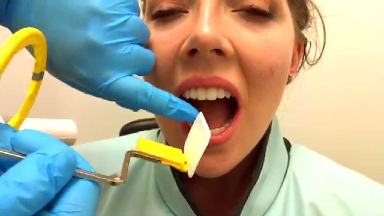
Students learn how to expose dental periapical x-ray film using the Rinn Paralleling Technique using the XCP film holder. Periapical X-ray images expor ting results and reading results. Single periapical radiographs are often made of individual teeth or groups of teeth to obtain information for treatment or diagnosis of localized diseases or abnormalities.
Parallel technique The image receptor is placed in a holder and placed in the mouth parallel to the longitudinal axis of the tooth under. The patient was positioned. 5 What is bisecting angle technique in dentistry.
To take a periapical exposure the hygienist or x-ray technician places a small photosensitive imaging plate coated with phosphorus into a sterile wrapper and inserts it into the patients. 3 What is an intraoral technique of exposing periapical images. The image receptor is placed in a holder and positioned in the mouth parallel to the long axis of the tooth under.
Size 2 Film is used for Anterior and Posterior X-rays when Bisecting. Paralleling Technique for Periapical X-rays The paralleling technique results in good quality x-rays with a minimum of distortion and is the most reliable technique for taking periapical x-rays. The X-ray tubehead is then aimed at right angles vertically.
Changes to the angulation of the X-ray beam in relation to the teeth and film can help diagnosis and treatment by producing images which provide additional information not alway. With this technique the film is placed parallel to the long axis of a tooth allowing the X-ray to be focused perpendicular to the long axis of the tooth. Periapical radiography is a commonly used intraoral imaging technique in radiology and may be a component of your radiologic examination.
When comparing the two periapical techniques the advantages of the bisecting. 4 Which technique is most often used when exposing a periapical image. Radiographic techniques 1.
Periapical images have been collected using the FONA X70 Intraoral X-rays machine and PSPIX Imaging Plates. Periapical X-rays. The extraoral periapical radiographic technique was performed for both maxillary and mandibular teeth using Newman and Friedman technique2.
A long cone is used to take x-rays with paralleling exposure techniques. Basically there are two techniques for taking periapical radiography. Occlusal X-rays show full tooth development.
A periapical x-ray or PA film will show one or two teeth in their entirety in one single image right from the crown of the tooth which is the part exposed in the mouth to the. The X-ray head is directed at right angles vertically and.
How To Take Periapical Radiographs Youtube

Periapical Radiography Pocket Dentistry

Periapical Radiography Pocket Dentistry

How Make Periapical X Ray

Periapical Radiography Pocket Dentistry

Periapical Radiography Pocket Dentistry

Periapical Radiography Pocket Dentistry

Periapical Radiography Pocket Dentistry
0 comments
Post a Comment